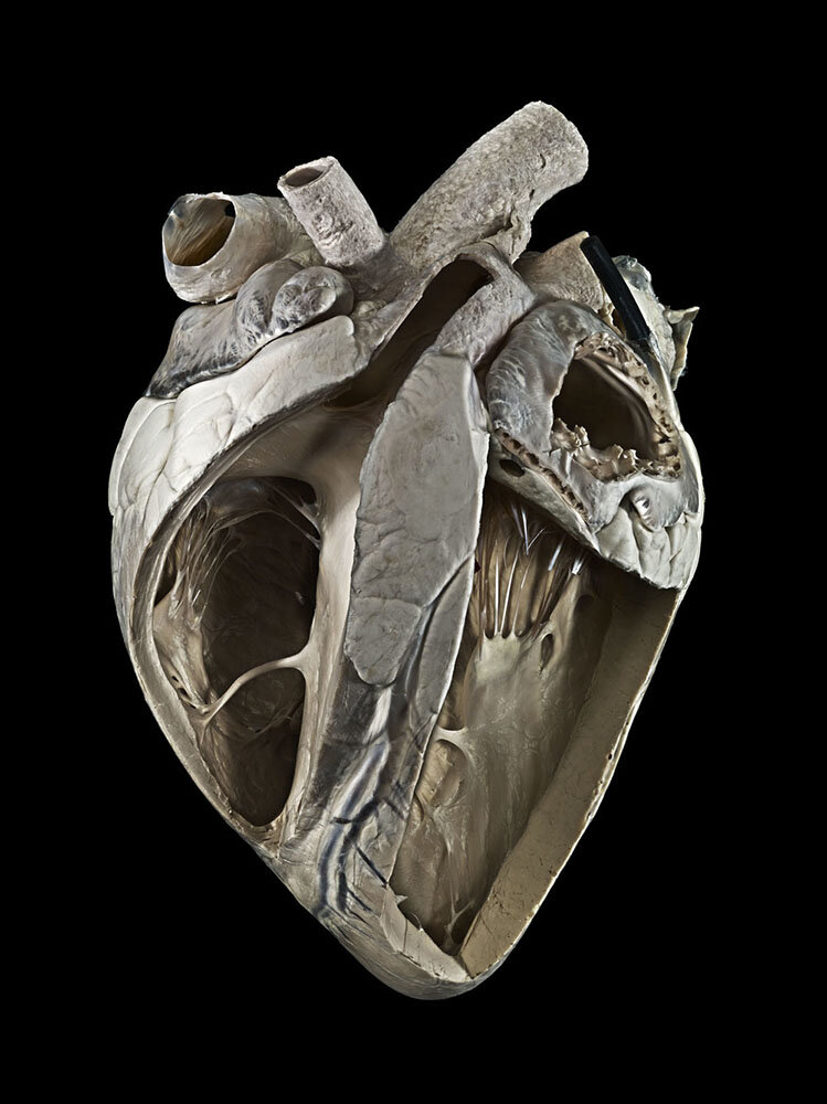
Bovine Heart
The image shows a bovine heart with left and right ventricles opened. The pot shows the moderator band (trebeculum septomarginalis) in the right ventricle and the ligamentum arteriosum at the pulmonary artery.

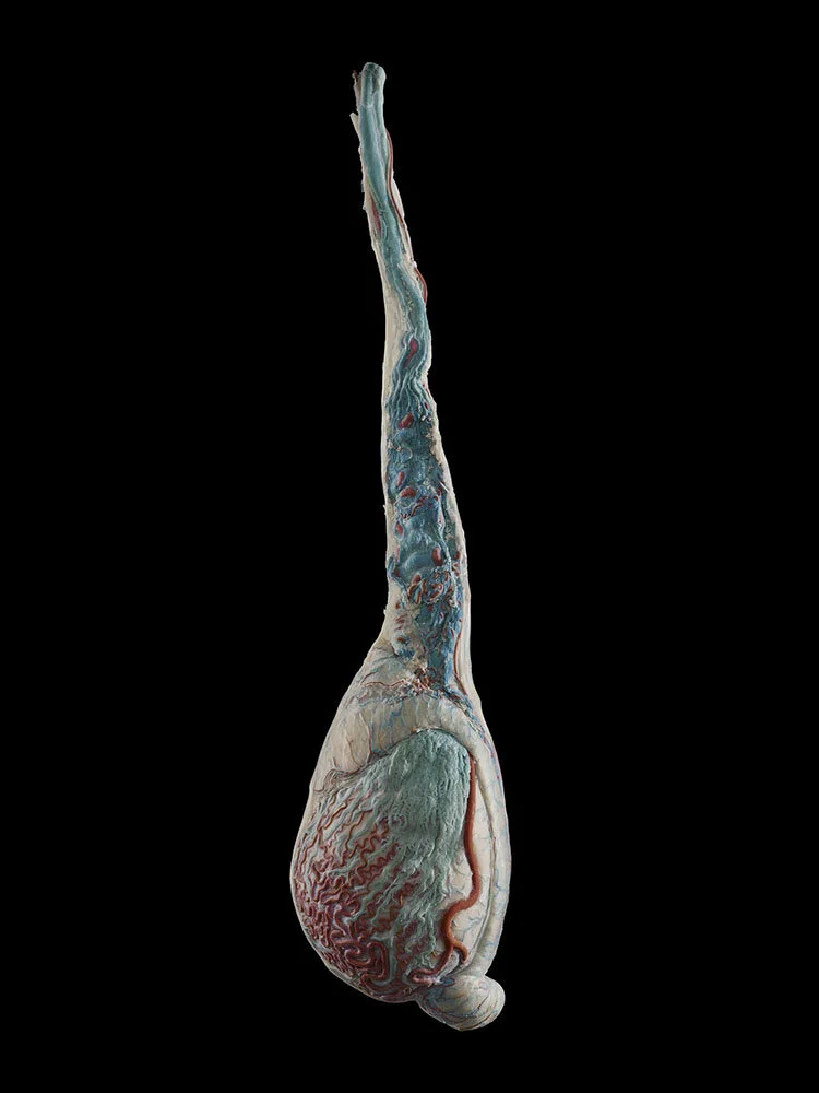
Pregnant uterus of a mare
The image shows a mare’s uterus with the foetus removed but still attached by the foetal membranes. The vast blood supply can be seen on the inner surface of the uterus.

Reticulum of a goat
This image shows the reticulum of a goat. The reticulum is one of the four chambers of the ruminant (cattle, goats, sheep) stomach and is characterised by the honeycomb pattern which lines the inside. Bacteria are present here which causes fermentation of the food material. The oesophagus can be seen entering the reticulum at the top of the picture. The opening in the centre leads to the omasum, another chamber of the stomach. A small part of the rumen, another chamber, is attached to the reticulum on the right. The lining changes here to small papillae, and resembles carpet.


Ovis Aries / domestic sheep
The reproductive organs of a ewe killed four days after copulation.
This is one of a series of preparations made by John Hunter.
Injected testis of a goat
The image shows an injected testis of a goat, showing the nature of the Pampiniform plexus heat exchange mechanism around the testicular artery.

Injected bull penis
The image shows an injected bull penis showing venous drainage and the sigmoid flexure common in ruminants and pigs.


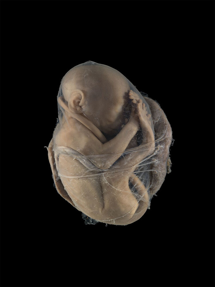
Foetus and discoid placenta of a monkey
This image shows a foetus of monkey enclosed in its foetal membranes. The discoid placenta, which supplies the foetus with nutrients, is attached behind it.

Form Follows Function / Anatomy Specimens
WINNER: 2015 Wellcome Image Awards
RVC shares anatomy images with Wellcome Trust
The eMedia Unit has been working with the Wellcome Trust to share images of anatomical specimens at the RVC. These range from historical dissections stored in formalin pots - brought alive by the latest in digital photography.
All images have been shot in the intern Anatomy Museum of the Royal Veterinary College, London.
Mick was honoured to be the recipient of the Wellcome Images Award 2015 for this series of work.
From the Wellcome Trust: ‘Our judges were unanimous in their decision, as James, Fergus and Sir Tim explain:
“As far as standout images go, the image of the horse’s uterus with the fetus was incredible and just sticks in my mind. It evokes many different emotions at once: it’s fascinating, sad, macabre, almost brutal. Yet the subject is also delicate, detailed and beautiful. The image shows us a large and magnificent creature reduced to this sad, fragile and half-formed creation, which I find very humbling.”
James Cutmore, Picture Editor of ‘BBC Focus’
“Those rear legs of a horse from an equine uterus – a compelling and haunting image.”
Fergus Walsh, Medical Correspondent for the BBC
“My favourite picture is of the equine uterus. Its presentation is hypnotic, like a Hieronymus Bosch painting… only it is real and truly marvellous.”
Sir Tim Smit, Founding Director of the Eden Project’
All images in this section are from:
Royal Veterinary College
Royal College of Surgeons of England
UCL Pathology Museum
Berliner Medizinhistorisches Museum der Charité